Home
/ Tendon Diagram Foot - Anatomy Of The Foot And Ankle Orthopaedia - 9 photos of the foot tendons and ligaments diagram.
Tendon Diagram Foot - Anatomy Of The Foot And Ankle Orthopaedia - 9 photos of the foot tendons and ligaments diagram.
Tendon Diagram Foot - Anatomy Of The Foot And Ankle Orthopaedia - 9 photos of the foot tendons and ligaments diagram.. When a muscle contracts, the tendon pulls on the bone causing the joint to move. Other important tendons in the foot include the tibialis posterior (posterior tibial tendon), which attaches the calf muscle to the bones on the inside of the foot and supports the arch of the foot, and the tibialis anterior (anterior tibial tendon), which runs from the outer tibia to the first metatarsal and surfaces of the median cuneiform tarsal, which allows for dorsiflexion—bringing the. Foot anatomy diagram, foot joint diagram, foot sprain diagram, foot tendons and ligaments pain, leg tendon diagram, peroneal tendonitis, foot, foot anatomy diagram, foot joint diagram, foot sprain diagram, foot tendons and ligaments pain, leg tendon diagram, peroneal tendonitis. Extensor foot tendons your extensor foot tendons connect muscle to bone on the top of your feet. To get started, all you need to do is click on the title of the.
When the muscles tighten (contract) arguably, the most important tendon is the achilles tendon, which allows the calf muscles to move. If you would like to learn all the parts of the foot structure, you have come to the right place. The achilles tendon is also called the calcaneal tendon. A tendon tear can be painful and make it hard to do any activities that require you to put weight on your foot. The contributors to this site are all board certified orthopaedic surgeons who specialize in treating patients with foot and ankle problems.
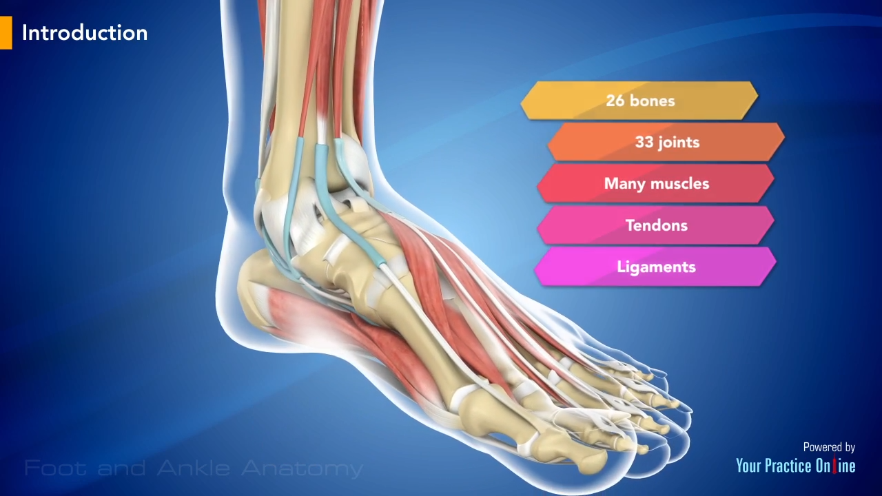
Foot And Ankle Anatomy Video Foot Ankle from www.ypo.education They are often associated with activities requiring increased pressure on the ball of the foot, such as running, basketball, football, golf, tennis and ballet. A foot tendon tear happens when one of the tendons in the foot is damaged from sudden injury or overuse. The tendons are thick bands that connect muscles to bones. A tendon is band of tissue made up of many fibers. The first foot pain diagram looks at the front and top of the foot, the second foot pain identifier looks underneath and at the back of the foot. Vector diagram of healthy foot and foot with gout. Swelling and bruising will occur at the site of injury. These tendons help your extensor muscles pull your foot upwards, which is necessary for walking.
A tendon is a band of tissue that connects a muscle to a bone.
The two peroneal tendons in the foot run side by side behind the outer ankle bone. They are often associated with activities requiring increased pressure on the ball of the foot, such as running, basketball, football, golf, tennis and ballet. The contributors to this site are all board certified orthopaedic surgeons who specialize in treating patients with foot and ankle problems. Front foot pain identifier this foot pain diagram shows common problems that cause pain on top of the foot at the front. More foot diagrams are on the following images. Muscles, tendons, and ligaments run along the surfaces of the feet, allowing the complex movements needed for motion and balance. If you would like to learn all the parts of the foot structure, you have come to the right place. Other tendons help to control the movements of the toes. A zone 1 injury involves an fdp tendon laceration distal to the fds insertion. The plantar ligaments are stronger than those on the dorsal side (figure 12 & 13). The first foot pain diagram looks at the front and top of the foot, the second foot pain identifier looks underneath and at the back of the foot. It connects muscle to bone. The achilles tendon is also called the calcaneal tendon.
Foot anatomy diagram, foot joint diagram, foot sprain diagram, foot tendons and ligaments pain, leg tendon diagram, peroneal tendonitis, foot, foot anatomy diagram, foot joint diagram, foot sprain diagram, foot tendons and ligaments pain, leg tendon diagram, peroneal tendonitis. Footeducation.com was created by orthopaedic surgeons to provide patients and medical providers with current and accurate information on foot and ankle conditions and their treatments. Attaches the calf muscles to the calcaneus, most important muscles for running, jumping, walking etc. Foot and ankle tendons that can get torn. The most common cause of tendonitis is overuse, which means a tendon is overly stretched and possibly experiencing a small degree of pulling apart or tearing.this occurs when there is an increase in activity, which can include anything from walking to participating in competitive sports.
Anatomy Of The Foot And Ankle Orthopaedia from orthopaedia.com The foot has a number of tendons. When a muscle contracts, the tendon pulls on the bone causing the joint to move. Allows the foot to be turned inward and also supports the arch of the foot. The first foot pain diagram looks at the front and top of the foot, the second foot pain identifier looks underneath and at the back of the foot. Allows the action of raising the foot. Extensor foot tendons your extensor foot tendons connect muscle to bone on the top of your feet. Match the corresponding numbers on the foot diagram below for a list of conditions that may be causing your foot and ankle pain. The achilles tendon is also called the calcaneal tendon.
Torn ligaments can occur following a range of physical activities from dancing to snowboarding, and several common symptoms can help identify a torn ligament as the cause of your foot pain.
Diagram of tendons in the foot. More foot diagrams are on the following images. Front foot pain identifier this foot pain diagram shows common problems that cause pain on top of the foot at the front. The muscles are located mainly in the sole of the foot and divided into a central (medial) group and a group on either side (lateral). The most popular tendon in our entire body is the achilles tendon, and it's also the most commonly injured. Allows the action of raising the foot. The first foot pain diagram looks at the front and top of the foot, the second foot pain identifier looks underneath and at the back of the foot. The tendons are thick bands that connect muscles to bones. Swelling and bruising will occur at the site of injury. The most common cause of tendonitis is overuse, which means a tendon is overly stretched and possibly experiencing a small degree of pulling apart or tearing.this occurs when there is an increase in activity, which can include anything from walking to participating in competitive sports. There are a number of tendons located in the foot and ankle all responsible for different ankle, foot and toe movements. When a muscle contracts, the tendon pulls on the bone causing the joint to move. The ankle serves as foundation, shock absorber and propulsion engine.
These ligaments prevent the joints of the midfoot from moving much, and as such provide considerable stability to the arch of the foot. In terms of mobility, the achilles tendon is one of the most important structures in the leg and foot. Here you can see the tendons that extend down the top of your foot toward your toes, allowing you to curl your toes upward if need be. A zone 1 injury involves an fdp tendon laceration distal to the fds insertion. A foot tendon tear happens when one of the tendons in the foot is damaged from sudden injury or overuse.
Https Encrypted Tbn0 Gstatic Com Images Q Tbn And9gctlfve Oxokw 2lm Mwksnhakj 5me7knvd6kbicurmicijxeb Usqp Cau from Footeducation.com was created by orthopaedic surgeons to provide patients and medical providers with current and accurate information on foot and ankle conditions and their treatments. Vector diagram of healthy foot and foot with gout. This tendon in the back of the calf and ankle connects the plantaris, calf, and soleus muscles. Front foot pain identifier this foot pain diagram shows common problems that cause pain on top of the foot at the front. Pain and tenderness are concentrated on the top, bottom or the sides of your foot near the arch. Torn ligaments can occur following a range of physical activities from dancing to snowboarding, and several common symptoms can help identify a torn ligament as the cause of your foot pain. Problems such as flat feet or high arches can create muscular. The muscles, tendons and ligaments.
Other important tendons in the foot include the tibialis posterior (posterior tibial tendon), which attaches the calf muscle to the bones on the inside of the foot and supports the arch of the foot, and the tibialis anterior (anterior tibial tendon), which runs from the outer tibia to the first metatarsal and surfaces of the median cuneiform tarsal, which allows for dorsiflexion—bringing the.
Diagram of tendons in the foot. A ligament is fibrous tissue that connects 2 or more bones together. The most common cause of tendonitis is overuse, which means a tendon is overly stretched and possibly experiencing a small degree of pulling apart or tearing.this occurs when there is an increase in activity, which can include anything from walking to participating in competitive sports. Here you can see the tendons that extend down the top of your foot toward your toes, allowing you to curl your toes upward if need be. The foot has a number of tendons. Torn ligaments can occur following a range of physical activities from dancing to snowboarding, and several common symptoms can help identify a torn ligament as the cause of your foot pain. Tendinitis can affect any tendon in the foot, and since there are so many of them, it may be difficult to tell what you have if you're in pain. It connects muscle to bone. The tendons are thick bands that connect muscles to bones. Other tendons help to control the movements of the toes. In addition, people with high arches are at risk for developing sesamoid problems. A tendon connects muscle to bone. A tear in either can come from trauma or repetitive stress.
The lisfranc joint complex is a series of ligaments that stabilize the tarsometatarsal joints tendon diagram. 9 photos of the foot tendons and ligaments diagram.